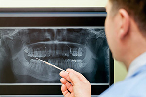
Our dental team promises to give our patients the best possible care by using digital X-rays in Yardley, Pennsylvania. Dr. Frank Prezioso and Dr. Robert Denmark can use the high-quality images produced by digital X-rays to give patients a quicker, more accurate response regarding the situation of their oral health. This method can help us better detect abnormalities in the mouth, including tumors, bone loss and tooth decay. Contact us today at 215-493-9325 for more information and to schedule your appointment with our dentists.
Safe, Fast and Detailed Imaging for Optimal Care
Digital X-rays are an essential diagnostic tool that allows our team to assess your oral health with precision and efficiency. Unlike traditional film X-rays, digital radiography utilizes advanced technology to capture high-quality images quickly and comfortably. This modern approach enhances your dental experience by reducing exposure to radiation, eliminating the need for chemical processing and providing instant results that can be reviewed during your appointment.
How Digital X-Rays Work
Digital X-rays use a small electronic sensor to capture detailed images of your teeth, gums and jawbone. These images are then transmitted to a computer, where they can be enlarged, enhanced and analyzed in real time. Because the process is immediate, we can quickly identify potential concerns and discuss treatment options without delay.
One of the primary advantages of digital X-rays is the significant reduction in radiation exposure. Compared to traditional film-based X-rays, digital radiography requires up to 90% less radiation, making it a safer option for patients of all ages. Additionally, because no chemicals are needed for film development, digital X-rays are an environmentally friendly choice.
Conditions Detected with Digital X-Rays
Digital X-rays provide valuable insight into various dental concerns that may not be visible during a standard examination. Our dentists may use digital radiography to diagnose and monitor conditions such as:
- Tooth Decay – Detects cavities between teeth and beneath fillings.
- Bone Loss – Identifies signs of periodontal disease or jawbone deterioration.
- Cysts and Abscesses – Reveals infections or fluid-filled pockets in the jaw.
- Impacted Teeth – Determines if wisdom teeth or other teeth are unable to erupt properly.
- Fractured Fillings – Helps assess damage to existing dental restorations.
- Developmental Abnormalities – Monitors tooth and jaw development in children and adolescents.
Benefits of Digital X-Rays
- Lower Radiation Exposure – Up to 90% less radiation than traditional X-rays.
- Instant Imaging – No waiting for film development, allowing for faster diagnosis and treatment.
- Enhanced Image Quality – Digital X-rays can be enlarged and adjusted for improved accuracy.
- Environmentally Friendly – No chemicals or film waste, reducing environmental impact.
- Easier Record-Keeping – Digital files can be stored, retrieved and shared effortlessly with specialists if needed.
Cone Beam Computed Tomography (CBCT)
We are proud to have a Planmeca ProMax 3D scanner in our office. It is a Cone Beam Computed Tomography (CBCT) imaging device that produces three-dimensional (3D) images of the teeth, jaw, and surrounding structures using a cone-shaped X-ray beam and computer processing, allowing for highly detailed visualization that traditional two-dimensional X-rays cannot provide. The scan is quick, painless and noninvasive.
Why is a CBCT Necessary At My Dental Appointment?
CBCT is widely used in dentistry. The images are used for detection of abscesses, cysts, tumors, fractured teeth and jaw or impacted teeth. In addition to its exceptional diagnostic value, the CBCT is used for dental implant planning to assess jawbone structure and optimal implant placement. It is used during root canal treatments to allow a 3D view of the tiny nerve canal that the doctor cleans and fills during a root canal treatment. The temporomandibular joint (TMJ) can also be more thoroughly evaluated with the 3D image of a CBCT.
Low Dose Imaging
The Planmeca Ultra Low Dose™ protocol can reduce CBCT radiation to levels lower than traditional 2D panoramic imaging, while still maintaining image quality. Exposure-control features on the unit automatically adjust radiation to the patient and imaging task, helping to keep doses as low as reasonably achievable (ALARA principle). Some QuickScan protocols provide high-resolution 3D images at radiation levels comparable to a 2D panorex, with no lingering radiation or side effects resulting in safer imaging for adults and children without sacrificing diagnostic detail. The Planmeca ProMaz 3D scanner, a CBCT, captures hundreds of image slices to build a full 3D model of dental anatomy, enabling highly detailed 3D images that are more accurate for diagnosis and treatment planning than 2D X-rays.
The ProMax 3D scanner offers a fast, painless and noninvasive experience. A complete 3D scan can take 10 to 30 seconds with the patient simply resting their jaw and biting on a bile block with their front teeth while the unit rotates around the outside of the patient’s mouth. This allows for more comfort because there is no biting on a large film or sensor inside the patient’s mouth. The open and spacious design of the machine helps prevent claustrophobic feelings during the imaging, resulting in less anxiety and more comfort during your dental visit.
Experience Advanced Dental Care
Digital X-rays play a crucial role in providing high-quality dental care by allowing for early detection and precise treatment planning. By incorporating this advanced technology, our practice ensures that you receive the safest, most efficient and most effective care possible. If you would like to learn more about digital X-rays and their benefits, contact our office today to schedule an appointment.
